- Product Details
- {{item.text}}
Quick Details
-
Brand Name:
-
DAWEI
-
Model Number:
-
RV-32B
-
Safety standard:
-
GB
-
Tabletop size:
-
1400*720mm, ±50mm
-
SID:
-
1000mm ±50mm
-
Imaging time:
-
≤1s
-
Focus:
-
1mm/2mm
-
Thermal capacity:
-
900KJ
-
Tube mA range:
-
10~400mA
-
Scintillator:
-
CSL
-
Imaging area:
-
430*430mm
-
Scanning matrix:
-
3072*3072 pixels
-
Pixel size:
-
140μm
Quick Details
-
Warranty:
-
2 years
-
After-sale Service:
-
Online technical support
-
Place of Origin:
-
Jiangsu, China
-
Brand Name:
-
DAWEI
-
Model Number:
-
RV-32B
-
Safety standard:
-
GB
-
Tabletop size:
-
1400*720mm, ±50mm
-
SID:
-
1000mm ±50mm
-
Imaging time:
-
≤1s
-
Focus:
-
1mm/2mm
-
Thermal capacity:
-
900KJ
-
Tube mA range:
-
10~400mA
-
Scintillator:
-
CSL
-
Imaging area:
-
430*430mm
-
Scanning matrix:
-
3072*3072 pixels
-
Pixel size:
-
140μm
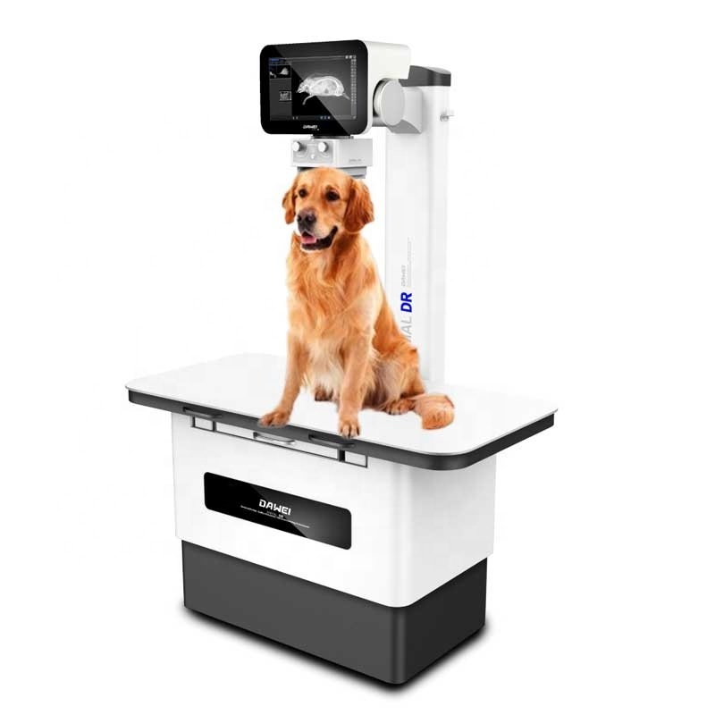
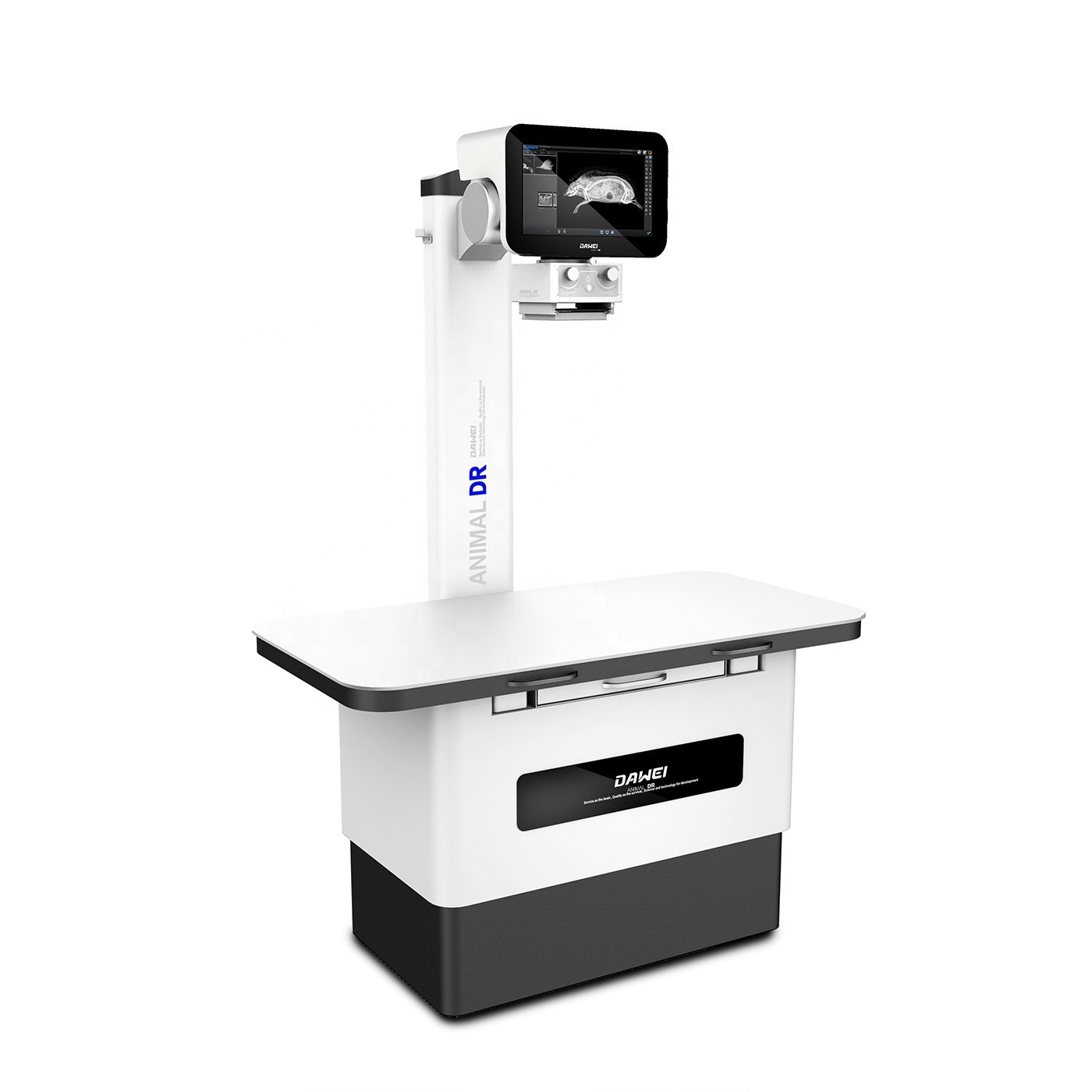
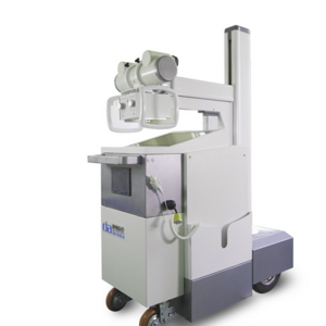
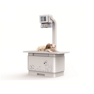
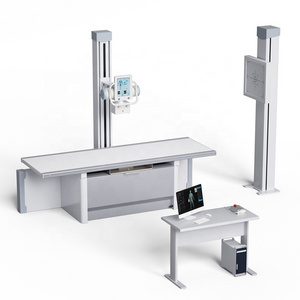
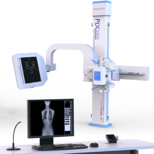
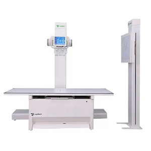
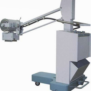
DAWEI pet dog diagnostic system veterinary digital radiography x ray machine
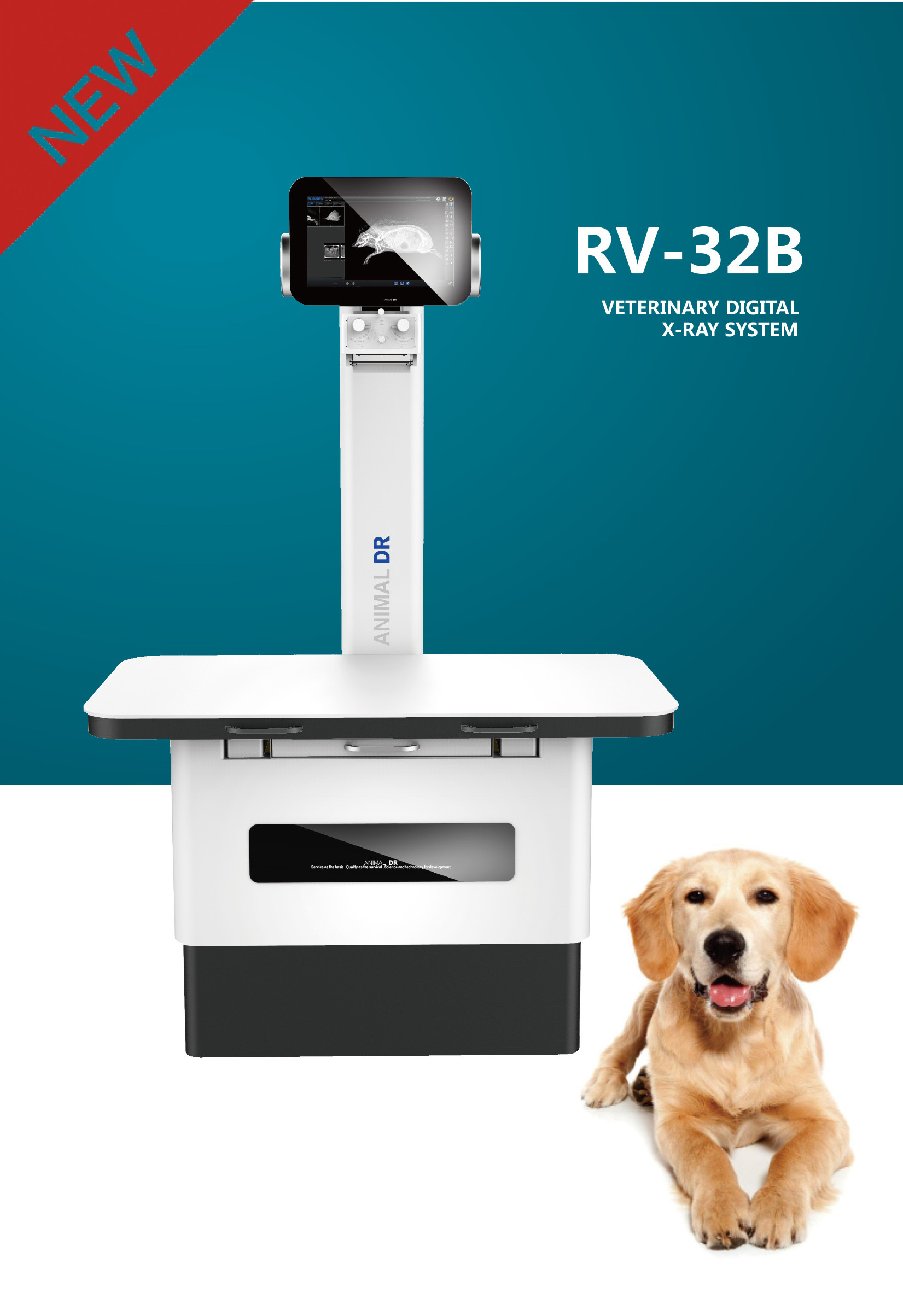
DAWEI Medical Radiography Veterinary X-ray Machine
·Tabletop size: 1400*720mm, + -50mm
·SID: 1000mm+ - 50mm
·Output: 32kw
·Tube mA range: 10~400mA
·Focus: 1mm/2mm
·Thermal capacity: 900KJ
·Scintillator: CSL
·Imaging area: 430*430mm
·Scanning matrix: 3072*3072 pixels
·Pixel size:140 µm
·Imaging time ≤1s
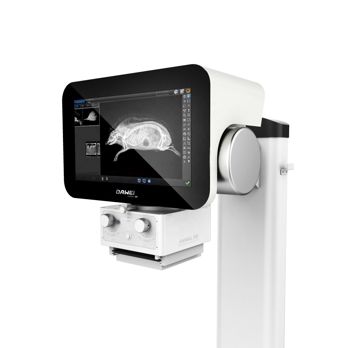
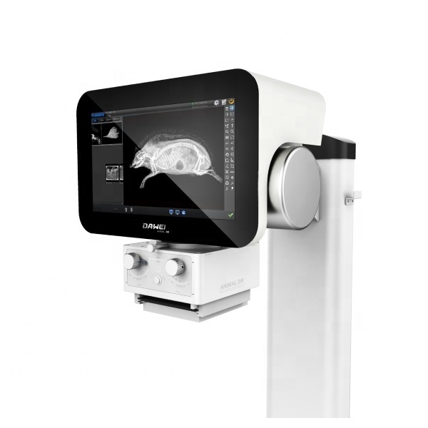
- 139um pixel size, spatial resolution of 3.7 lp/mm, pixel matrix 3072*3072 px, megapixel imaging.
-
Tablet's unique automatic exposure detection function, referred to as AED.
-
Amorphous silicon cesium iodide
: has high X-ray conversion efficiency, conversion speed and quantum detection efficiency DQE.
- Complete shooting within
3s
.
-
16-bit large dynamic range
-
High stability: The flat-panel detectors used by Dawei RV-32A&RV-32B basically have no loss of connection; the use of environmental temperature (5℃~35℃) and humidity (10%~75%)
- Dual focus: small focus 1.0mm, large focus 2.0mm, to achieve the best balance of image resolution and loading power.
-
High heat capacity: The heat capacity of the tube is 140kHU, which achieves more stable continuous exposure performance and better meets the requirements of continuous work.
-
Maximum tube current: 340mA for small focus and 570mA for large focus, greatly improving photography conditions. Suitable for a variety of animal body types and body positions to meet the loading conditions of a variety of special photography.
- Intelligent adjustment of exposure parameters
- Real-time synchronization control with workstation
- Real-time display of captured images
- 15.6-inch IPS capacitive touch LCD screen
Software
This system is a set of medical software developed by our company for the purpose of facilitating doctors' diagnosis and patient consultation. Using this software, doctors can complete inspection items such as the collection, transmission and film printing of patient X-ray examination pictures. The multiple image processing methods built into the software also greatly facilitate the doctor's consultation, which can greatly improve the speed and accuracy of consultation. The specific functions are as follows:
✸Examination registration management: register patient information, store it in the database, and associate it with the image of the patient examination.
✸ Filming inspection: According to the registered inspection site, the filming inspection can be carried out, and the position can be added or deleted at any time, and the filming dose can be adjusted.
✸Diagnosis assistance: Enhance DR images, and provide a series of auxiliary diagnosis tools such as image adjustment, cropping, and marking.
✸Case management: patient cases and examination images are stored in a database for browsing at any time.
✸System docking: Using standard DICOM 3.0 communication protocol, it can easily connect to hospital HIS, RIS, PACS and other systems, and connect to DICOM film printers.
✸Film printing: supports local printing and DICOM network printing.
✸Case management: diagnosis report: reports can be set according to the needs of hospitals or departments, and the report layout can be edited.
|
Tabletop size
|
1400*720mm, ±50mm
|
|
SID
|
1000mm ± 50mm
|
|
Output
|
32kw
|
|
Tube mA range
|
10~400mA
|
|
Focus
|
1mm/2mm
|
|
Thermal capacity
|
900KJ
|
|
Scintillator
|
CSL
|
|
Imaging area
|
430*430mm
|
|
Scanning matrix
|
3072*3072 pixels
|
|
Pixel size
|
140 µm
|
|
Imaging time
|
≤1s
|
Imaging
Hot Searches
