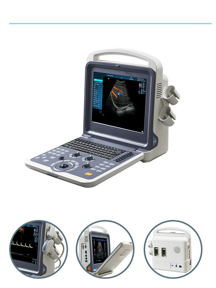
- Product Details
- {{item.text}}
Quick Details
-
Brand Name:
-
AMIS
-
Model Number:
-
AM-K0
-
Quality Certification:
-
ce,ios
-
Display mode:
-
2B, 4B, M, B/M, B/C, B/D, B/C/D, B/CFM/D, PDI
-
Display:
-
15 inch LED Monitor
-
Language:
-
multi-language
-
storage:
-
120G memory
-
Probe elements:
-
80
-
Gray:
-
256
-
Image storage format:
-
BMP, JPEG, PNG, DICOM(Option)
-
Cine loop:
-
1200 frames
Quick Details
-
Warranty:
-
2 years
-
After-sale Service:
-
Online technical support
-
Place of Origin:
-
Jiangsu, China
-
Brand Name:
-
AMIS
-
Model Number:
-
AM-K0
-
Quality Certification:
-
ce,ios
-
Display mode:
-
2B, 4B, M, B/M, B/C, B/D, B/C/D, B/CFM/D, PDI
-
Display:
-
15 inch LED Monitor
-
Language:
-
multi-language
-
storage:
-
120G memory
-
Probe elements:
-
80
-
Gray:
-
256
-
Image storage format:
-
BMP, JPEG, PNG, DICOM(Option)
-
Cine loop:
-
1200 frames
Product Display

Smart design
High Resolution Imaging System
Easy-operation Ergonomic Design
Better Optimize Image Quality
Smart and Light -Weight Design
Probes information

3.5Mhz abdominal probe
2.0, 3.0, 3.5, 4.0, 5.5Mhz

7.5Mhz linear probe
6.0, 6.5, 7.5, 10.0, 12.0Mhz

6.5Mhz transvaginal probe
5.0, 6.0, 6.5, 7.5, 9.0Mhz
Product Details
|
Displaying mode
|
B,B/B,4B,B/M,M, B/C,B/C/D,B/D, duplex, triplex, CFM, PW
|
|
|
Full-digital beam forming, dynamic filter, dynamic real time receiving focusing, spectral processing, CFM processing, real-time
dynamic focusing, dynamic aperture in all fields |
|
Image processing
|
THI
|
|
|
Storage: 16G
|
|
|
Power adjustable
|
|
|
Smoothing function
|
|
|
Edge enhancement
|
|
|
One-key optimization
|
|
|
Image conversion
|
|
|
Wall filter adjustable
|
|
|
Base line adjustable
|
|
|
PRF adjustable
|
|
|
AIO-Auto image optimization
|
|
|
IZoom: Instant full screen image
|
|
|
I-Image: intelligent optimization
|
|
|
MBF: Multi Beam Former
|
|
|
SA: Synthetic Aperture Ultrasonic imaging
|
|
|
Iclear: Speckle Noise Reduction
|
|
|
CDF: Contunuous Dynamic Focusing
|
|
General measurement
|
Normal, MSK, ABD, OB, Pelvic,Urology, Cardiac, Small Parts, Vascular
|
|
|
Volume, V3L, STD_S, Area Trace,Mtime, MHR, D Time, DV, D Common, D Auto, Area, Angle, CrossLine, STD D, ParalleLine, Mdist,MV, D
HR, DA, D Trace |
|
ABD packages
|
ABD, Aorta, R_Kidney & L_Kidney, Bladder, Prostate
|
|
OB Packages
|
Early_OB, Rt-Ovary, Lt-Ovary, Uterus, Fetal_Biome, Long_Bones, AFI
|
|
Pelvic Packages
|
Uterus, Rt/Lt - Ovary, Rt/Lt-Follicle,
|
|
|
|
|
Urology packages
|
Rt/Lt- kidney measurement, Bladder, Prostate,Rt/Lt_Testicle
|
|
|
|
|
Small Parts
|
Rt/Lt_Thyroid, Rt/Lt_Testicle, Vessel, Breast
|
|
Vascular
|
Stenosis D, senosis A, Intima, Arterial, Venous
|
|
MSK
|
Distance, Area, Hip_Angle
|
|
Scanning depth
|
≥260mm
|
|
Probe elements
|
80
|
|
Gray
|
256
|
|
Cine loop
|
Automatically & manually
|
|
Image storage format
|
BMP, JPEG, PNG, DICOM(Option)
|
|
|
|
|
Input/output ports
|
Video Port, S-Video Port, Remote Port, LAN1/2 Port, VGA
|
|
Standard Configuration
|
Main unit, 12 inch LED monitor, 3.5Mhz convex probe, 7.5Mhz linear probe, 2 Probe connectors, User’s Manual, hard disk (SSD)
|
|
Options
|
6.5Mhz Transvaginal Probe, Trolley, Printers,Aluminum case
|
|
Applicable Printers
|
HP Laser Jet P2035
|
|
|
HP Laser Jet 1022
|
|
|
HP Laser Jet 1020
|
|
|
SONY UP-D898MD
|
|
|
SONY UP-X898MD
|
|
|
SONY UP-897MD
|
|
|
EPSON L130
|
|
|
EPSON L805
|
Detail Image
Application
PW : Pulse Wave Doppler
The launch and reception of Ultrasonic pulse wave are processed by one probe,and start to receive echo signal after delay of
schedule time.
schedule time.
Pseudo
Pseudo color processing will change the gray level image to color image.Pseudo color makes the users distinguish the different
organs issue.KO's Pseudo color is more than 15 kinds of different color.
organs issue.KO's Pseudo color is more than 15 kinds of different color.
CF : Color Flow Mode
Double ultrasonic scanner system which could show B mode image and Doppler blood flow data(ie.blood flow direction,speed,velocity
dispersion) simultaneously.
dispersion) simultaneously.
THI : Tissue Harmonic lmaging
Tissue Harmonic Imaging,which avoids many of the near-field artifacts that affect fundamental imaging. And have the advantage of
good signal-noise ratio, better spatial resolution,enhance tissue contrast, Improve deep tissue echo information.
good signal-noise ratio, better spatial resolution,enhance tissue contrast, Improve deep tissue echo information.
Recommend Products
Why Choose Us
Hot Searches


