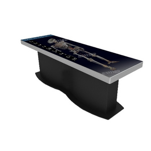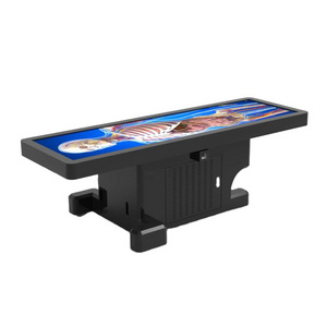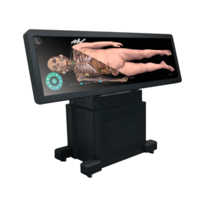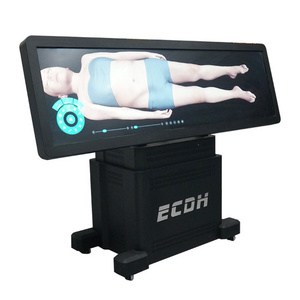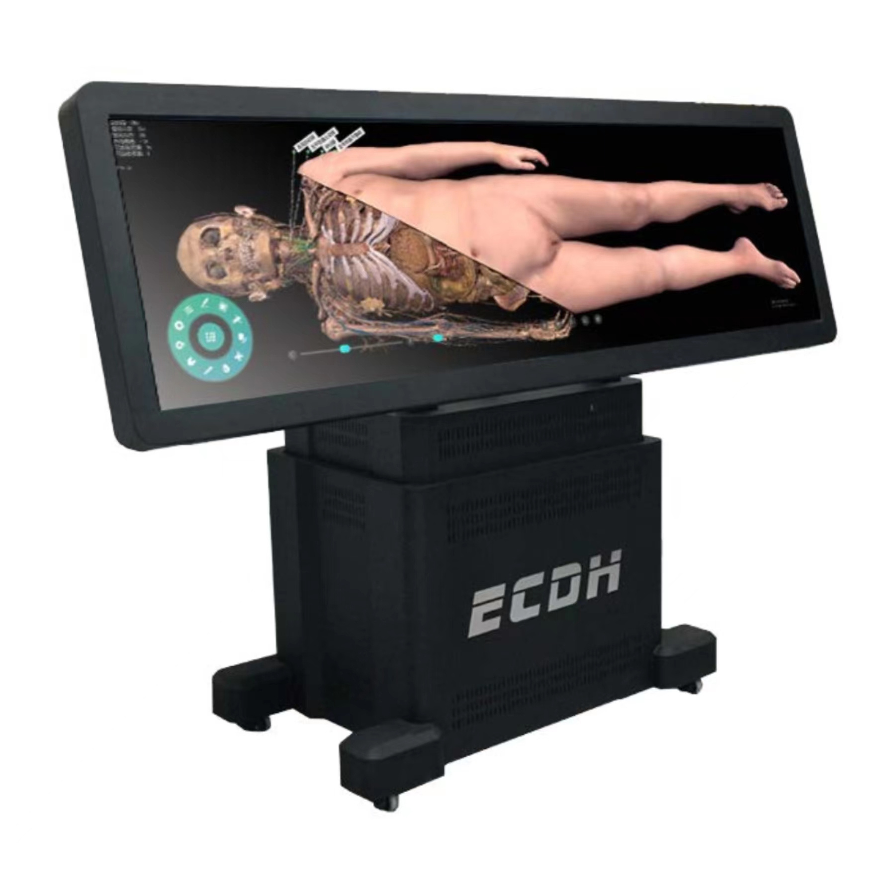
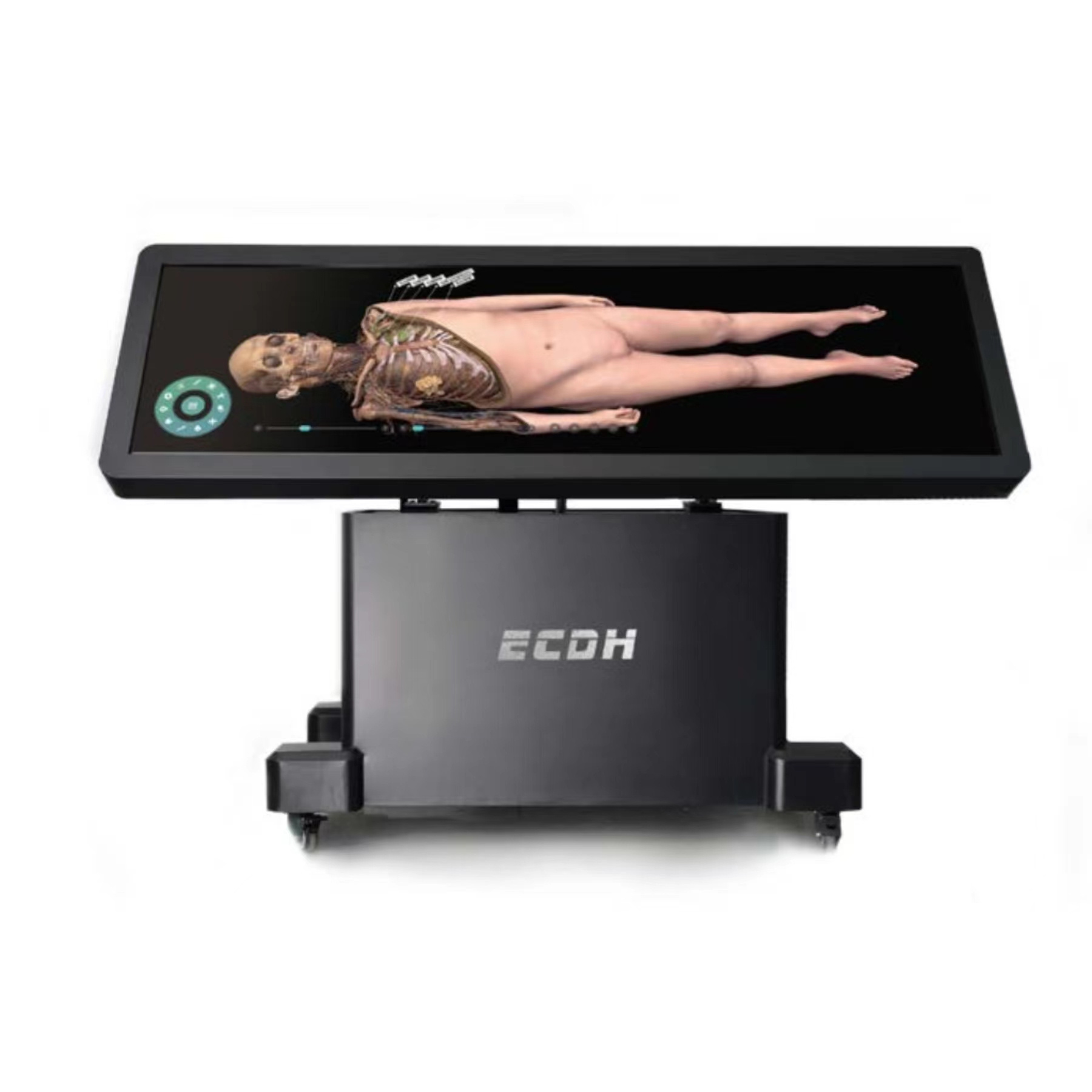

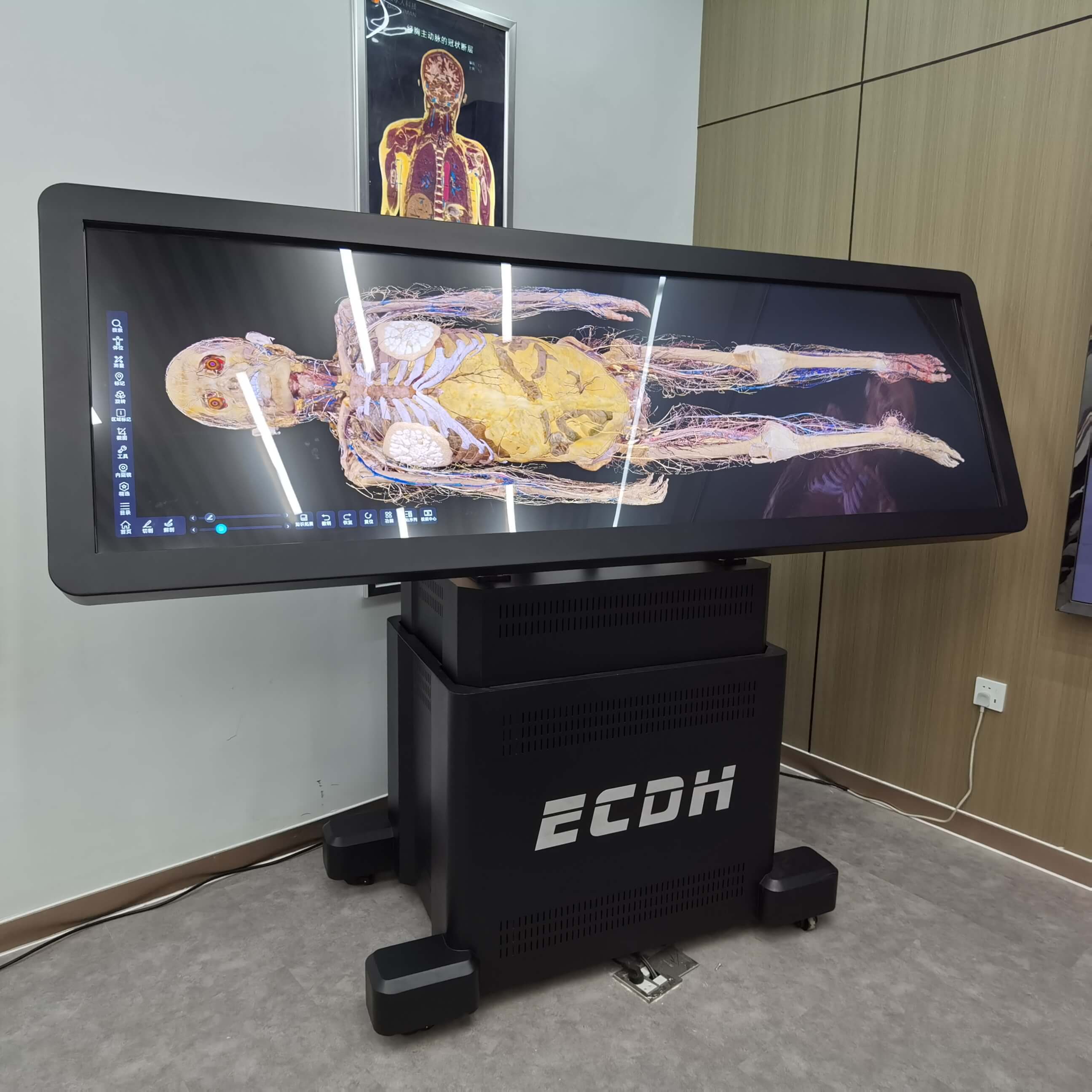
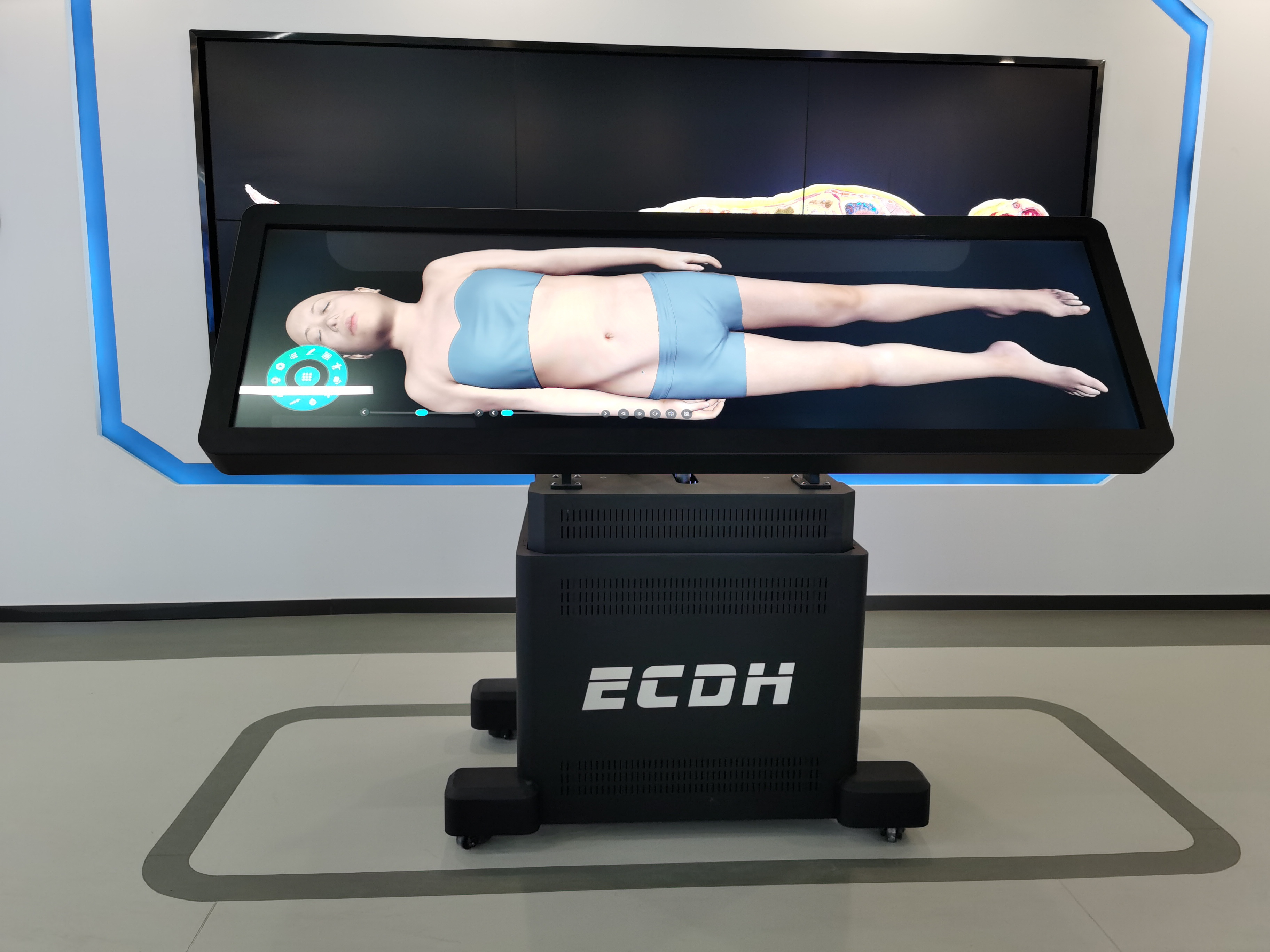
Digihuman Virtual Anatomage Tableis a set of anatomy equipment creating the virtual structure of the human body with the digital 3Deconstruction technology. The anatomy contents are displayed with a natural structure, the scale of a real person, touch screen interaction, the angle of view in the supine position, the conversion of horizontal and vertical screens, and both Chinese and English.
Through the technical processing of sequence images of sections of the human body with ultra-high precision, the structure of the human body can be drawn in real-time with ultra-high precision and a lot of data. The touch control operation of the Anatomage Table can realize the cutting and observation of the structure of the human body in different directions and angles at different lavers, The knowledge of human anatomical anatomy. and sectional anatomy is integrated. With the classical section as the axis, the virtual cutter is used for the continuous display. By constructing the virtual anatomy combination model, it helps the observer to develop the concept of the 3D space structure.
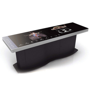
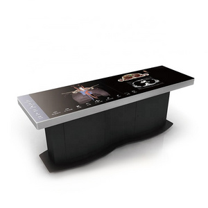
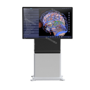
The system integrates many kinds f medical visualization resources and human-computer interaction techniques. The UHD realistic anatomy system of the human body, the Digihuman Anatomy System, the user interaction management system, and other systems are integrated to construct the virtual training platform. With features such as high-precision realistic anatomical structure, the high-performance anatomy teaching tool, and the interactive touch control operation, it can help users to learn about the macroscopic microscopic, normal, and sick 3D structure.
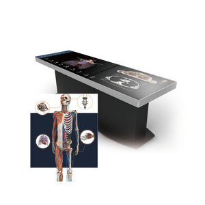
The system contains five parts: "Human anatomy","Digital human anatomy system", "Slice library" , " Clinical cases" and “ Embryology”.
Human Anatomy
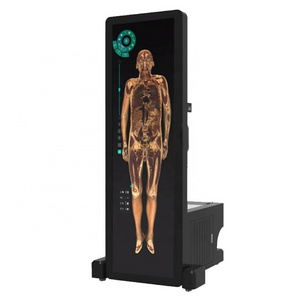
The 3D structure of the human body in the human anatomy module is digitally restored from male data (17000+ total layers of cross-section; resolution 13700*6340) and female data (16000+ total layers of cross-section; resolution 12000*5700).
Able to perform virtual anatomy operations through it.
Anatomy System
The system contains a
wealth of learning resources:
more than 3000
3D structures,
more than 3000 sectional images, more than
1700 images of CT/MRI, more than 100 teaching videos and more than 1800 exercises.
Through the "Digihuman Anatomy Teaching System", teachers can operate professional anatomy teaching soft-ware through the touch pad. Students can watch stereo human anatomy structures by wearing 3D glasses, which can be viewed in an open, autonomous and
interactive virtual environment Conduct an efficient, safe and eco-nomical teaching process.
Embryology
The system is a collection of videos, animations, 3D models and courseware as an integrated teaching platform.
The system is categorized in three modules on early human embryogenesis (General), the genesis of human embryonic organ systems (Monographs) and congenital malformations, each of which has the following teaching resources: preface, general video, knowledge points and exercise bank.
Slice library
The section library module contains at least 395 digital sections of histology and 780 digital sections of pathology.
The slice library supports touch or mouse to simulate the operation under the mirror. One-click 4X, 10X, 20X, 40X objective lens magnification adjustment, also can pan to adjust the observation position, one click to select the history of browsing sections or favorite sections.
Clinical cases
The clinical case module contains the number of real clinical cases not less than 180. It can display the disease name, basic information, chief complaint, image performance and diagnosis of the current case. The system provides window adjustment for CT/MRI images, and the window width and window position can be manually adjusted according to different parts so that users can quickly view different image contents.