- Product Details
- {{item.text}}
Quick Details
-
Brand Name:
-
FORERMED
-
Product name:
-
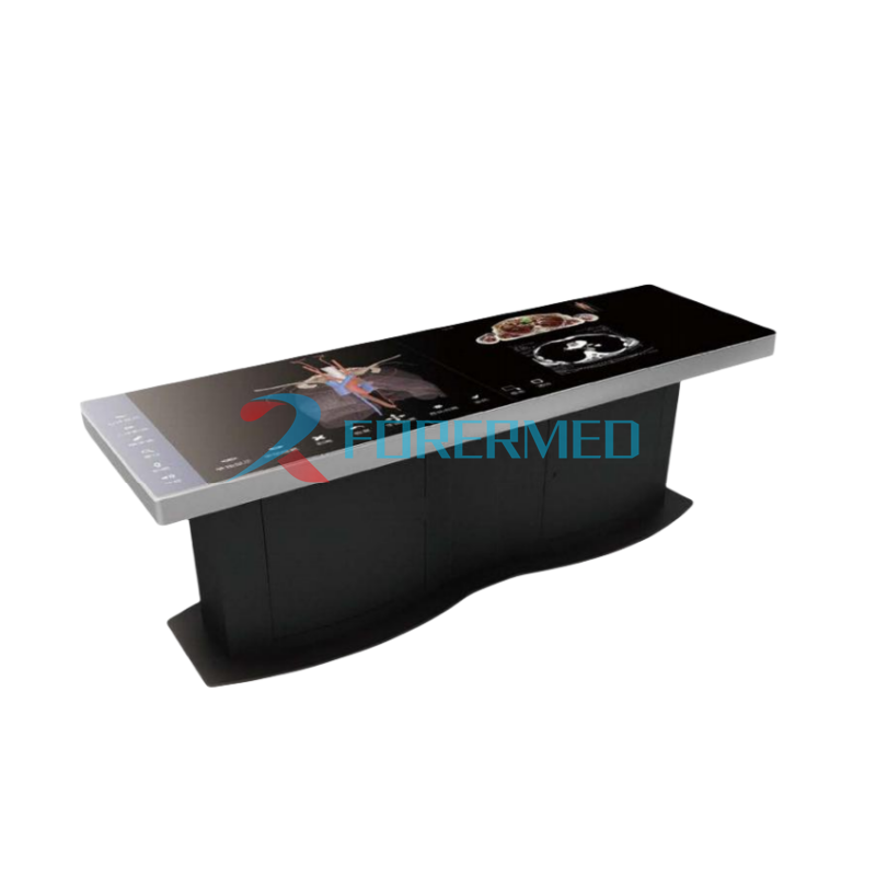
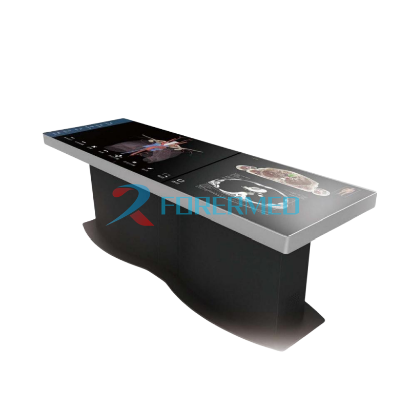
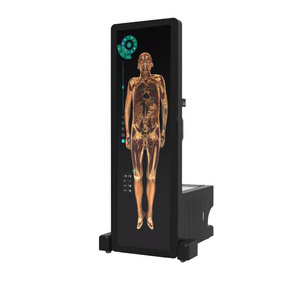
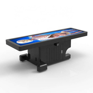
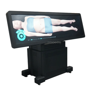
Digihuman Virtual Anatomy Table
-
Application:
-
Medical School
-
Display:
-
2 sets of 47 inch splicing screens with 3.5mm splicing seam
-
Resolution:
-
3480*1080
-
Brightness:
-
500
-
Contrast ratio:
-
1100:1
-
Elevation:
-
175
-
Weight:
-
185kg
-
Dimensions:
-
2260*707*750mm
Quick Details
-
Type:
-
Anatomical Model
-
Model Number:
-
YJ-DHA88B
-
Place of Origin:
-
Henan, China
-
Brand Name:
-
FORERMED
-
Product name:
-
Digihuman Virtual Anatomy Table
-
Application:
-
Medical School
-
Display:
-
2 sets of 47 inch splicing screens with 3.5mm splicing seam
-
Resolution:
-
3480*1080
-
Brightness:
-
500
-
Contrast ratio:
-
1100:1
-
Elevation:
-
175
-
Weight:
-
185kg
-
Dimensions:
-
2260*707*750mm
Product Description
Product Description
1 The digital human anatomy system based on the 3D reconstruction of continuous real sectional images.
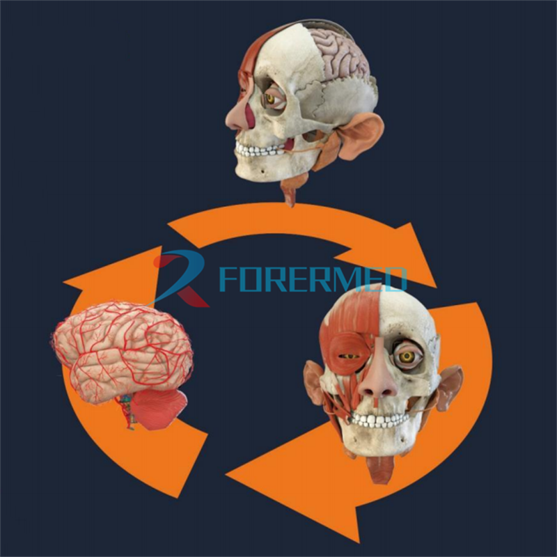
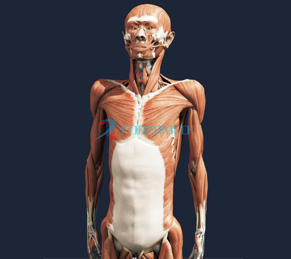
The system is developed with continuous real sectional images of human specimen and more than 5000 3D reconstructed anatomical structures.
2 Full-featured digital anatomy teaching system.
The system can display all the human organs and tissues in completely realistic 30 model. Each structure is set with English names and English pronunciation, and all the key structures are marked with d etailed annotation and corresponding textual interpretation. The anatomy structures can be rotated and viewed at any angle, The system setting functions including background switching, labeling, separation, transparency, dyeing, stripping, searching, pronunciation, freehand drawing and stereotaxic display etc. it can strengthen the vitality, interestand intuition of anatomy teaching.
3 Student autonomous learning system.
The system covers anatomy teaching contents. Corresponding CT and magnetic resonance images are arranged on the basis of the image of the section specimen. Also provide teaching micro-course video and a large number of digital exercises.
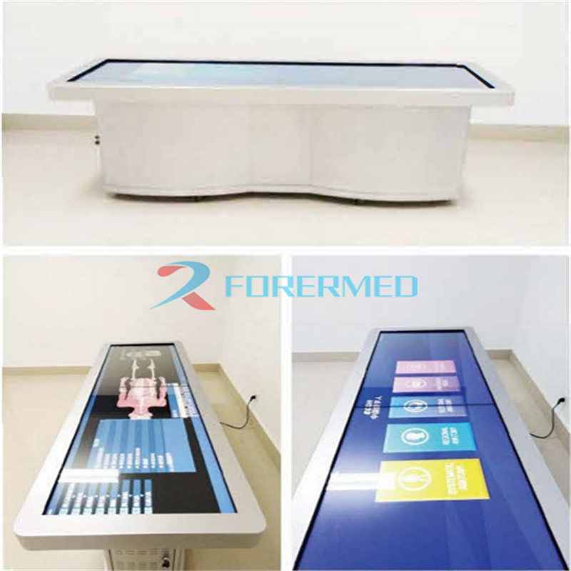
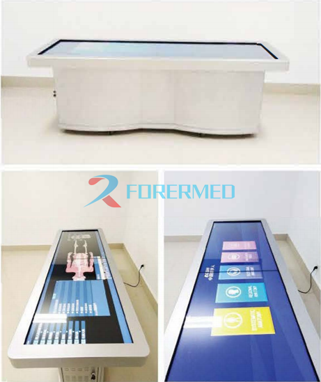
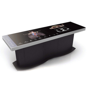
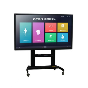
4 Simple and quick full touch operating system.
The system uses full touch operation interface with a 86/55-inch multi-touch system embedded, which has simple
structure and beautiful appearance. It can power up to work without any software installation and debugging procedures.
The system is developed with continuous real sectional images of human specimen and more than 5000 3D reconstructed anatomical structures.
2 Full-featured digital anatomy teaching system.
The system can display all the human organs and tissues in completely realistic 30 model. Each structure is set with English names and English pronunciation, and all the key structures are marked with d etailed annotation and corresponding textual interpretation. The anatomy structures can be rotated and viewed at any angle, The system setting functions including background switching, labeling, separation, transparency, dyeing, stripping, searching, pronunciation, freehand drawing and stereotaxic display etc. it can strengthen the vitality, interestand intuition of anatomy teaching.
3 Student autonomous learning system.
The system covers anatomy teaching contents. Corresponding CT and magnetic resonance images are arranged on the basis of the image of the section specimen. Also provide teaching micro-course video and a large number of digital exercises.
4 Simple and quick full touch operating system.
The system uses full touch operation interface with a 86/55-inch multi-touch system embedded, which has simple
structure and beautiful appearance. It can power up to work without any software installation and debugging procedures.
Technical Parameter
|
Product Specification
|
|
|
|
Dimensions:
|
length 2260mm,width 707mm,height 750mm
|
|
|
Weight:
|
185Kg
|
|
|
Display:
|
2 sets of 47 inch splicing screens with 3.5mm splicing seam
|
|
|
Resolution Ratio:
|
3480*1080
|
|
|
Brightness:
|
500 cd/㎡
|
|
|
Contrast Ratio:
|
1100:01:00
|
|
|
Visual Elevation:
|
175°
|
|
Features
Accurate Data
Clear lmage
Section Precision
0.1-1 mm
The system is developed using continuous transverse sectional images of human specimens. The section precision for men and women was 0.1-1mm and 0.1-0.5mm, respectively, and the thickness is unequal. In the parts of head and chest, the layer spacing is up to 0.1mm because they need to be displayed finely
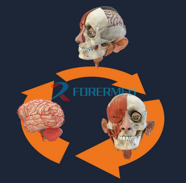
Stereoscopic Observation
The digital human can be rotated at any angle and arbitrarily zoom in and out. It can be observed in all directions from the perspective of looking up and looking down.
The structures will be more visual and intuitive contrast with the models and specimens.
Intra-pulmonary pipe
Fiber Bundles in the brainstem
Lateral view of the skull
Arbitrary angle rotation
Arbitrary zoom in and out
3D stereoscopic observation
3D Structurs
Fine and Realistic
More than 6000 fine and realistic reconstructed anatomical structures were embedded in Digital Human Anatomy System, which can provide a lot of material for anatomy teaching.
Rich in Functions Easy To Operate
The system has designed a variety of quick and convenient functions, including background switching, labeling, separation, transparency, dyeing, stripping, searching, bilingual pronunciation, freehand drawing and stereotaxic display
et al.
Note: The stereoscopic display function requires hardware support
Teaching Application
Systematic Anatomy
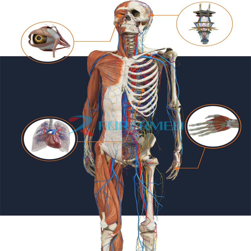
The three-dimensional structures are obtained by 3D reconstruction of real human cross-sectional data. Their position and shape are consistent with the original data.The structures are divided into nine systems.And the three-dimensional morphology of more than 6000 anatomical structures can be displayed.
Nine Systems
Locomotor System; Angiology System; Alimentary System; Sensory System Espiratory System; Nervous System; Urinary System; Endocrine System; Reproductive System
Sectional Anatomy
It’s easy to obtain sectional images of any section.
Using the highlighting function, the sectional structures can be identified, their Chinese and English names can be obtained quickly and their positions and shapes can be showed in the three- dimensional human body. Which can provide real specimens and imaging images for studentslearning sectional anatomy.
Regional Anatomy
For the teaching of Regional Anatomy, teachers can display the structures from superficial layer to deep layer using the digital human body with stripping and perspective functions. The students are able to build local hierarchical concepts and know the adjacent relationships of the structures even in the classroom.The Digital Human Anatomy System includes a large number of regional anatomy teaching videos to facilitate teaching and students' self-study
Surface Anatomy
The surface projection of nerves, blood vessels and other structures can be realized with the transparent function of the digital human.
Hot Selling
Why Choose Us


Hot Searches
