
- Product Details
- {{item.text}}
Quick Details
-
Brand Name:
-
ECDH
-
scanning space:
-
1mm-3mm
-
3D structure resolution:
-
4K
-
Display:
-
1960mm*1147
-
Resolution:
-
3840*2160
-
Size:
-
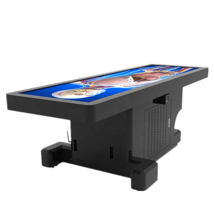
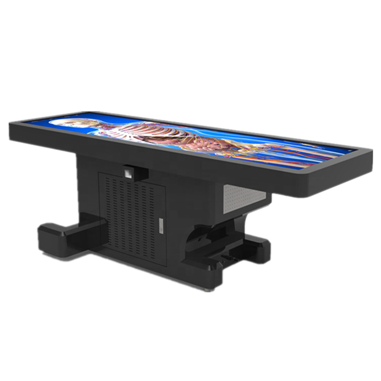
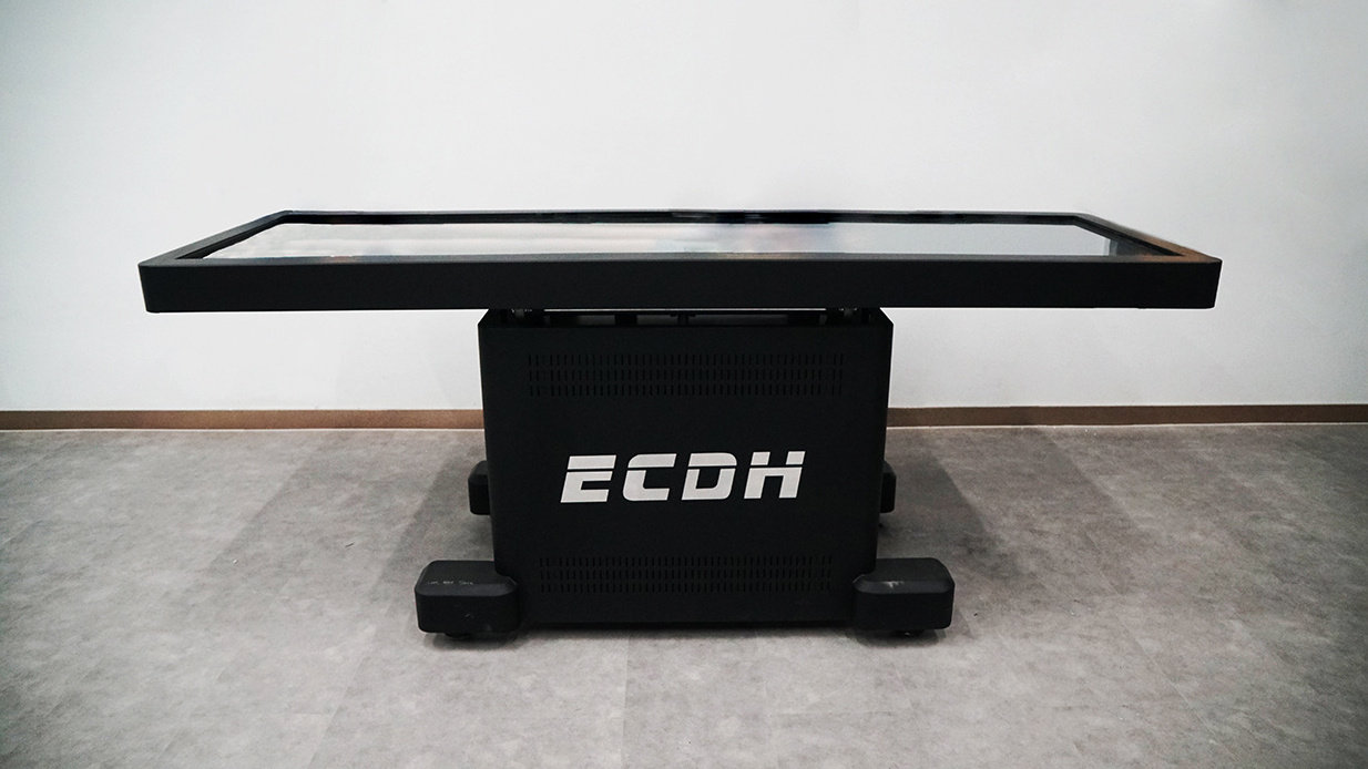
L:2260mm; W:707mm; H:750mm
-
Weight:
-
85Kg
-
Brightness:
-
850cd/m2
Quick Details
-
Type:
-
Anatomical Model
-
Model Number:
-
ECDH-II 88
-
Place of Origin:
-
Shandong, China
-
Brand Name:
-
ECDH
-
scanning space:
-
1mm-3mm
-
3D structure resolution:
-
4K
-
Display:
-
1960mm*1147
-
Resolution:
-
3840*2160
-
Size:
-
L:2260mm; W:707mm; H:750mm
-
Weight:
-
85Kg
-
Brightness:
-
850cd/m2
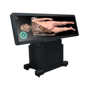
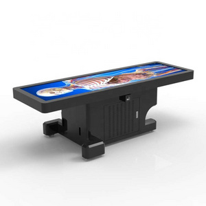
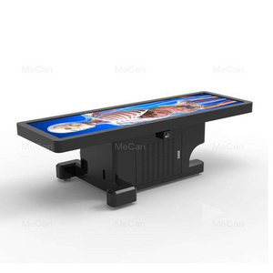
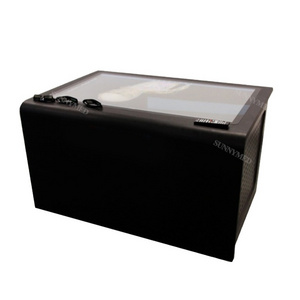
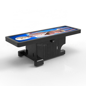
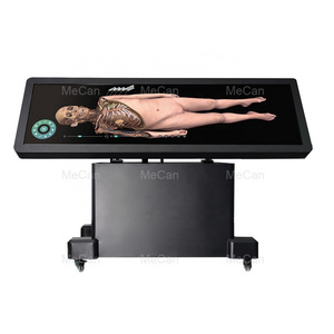
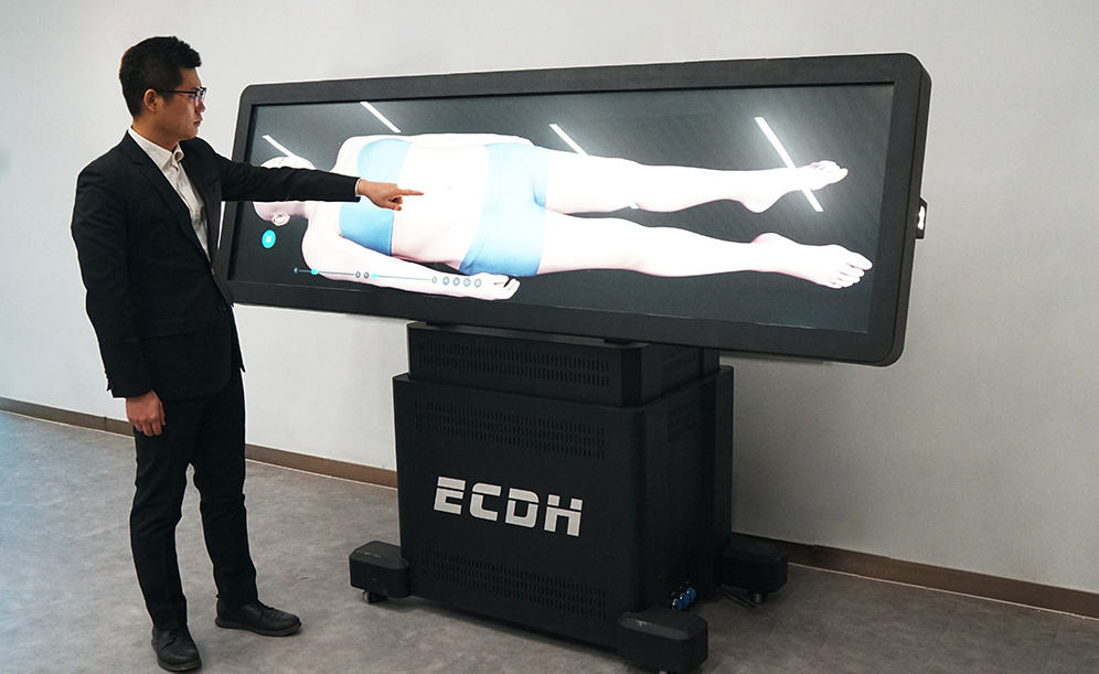
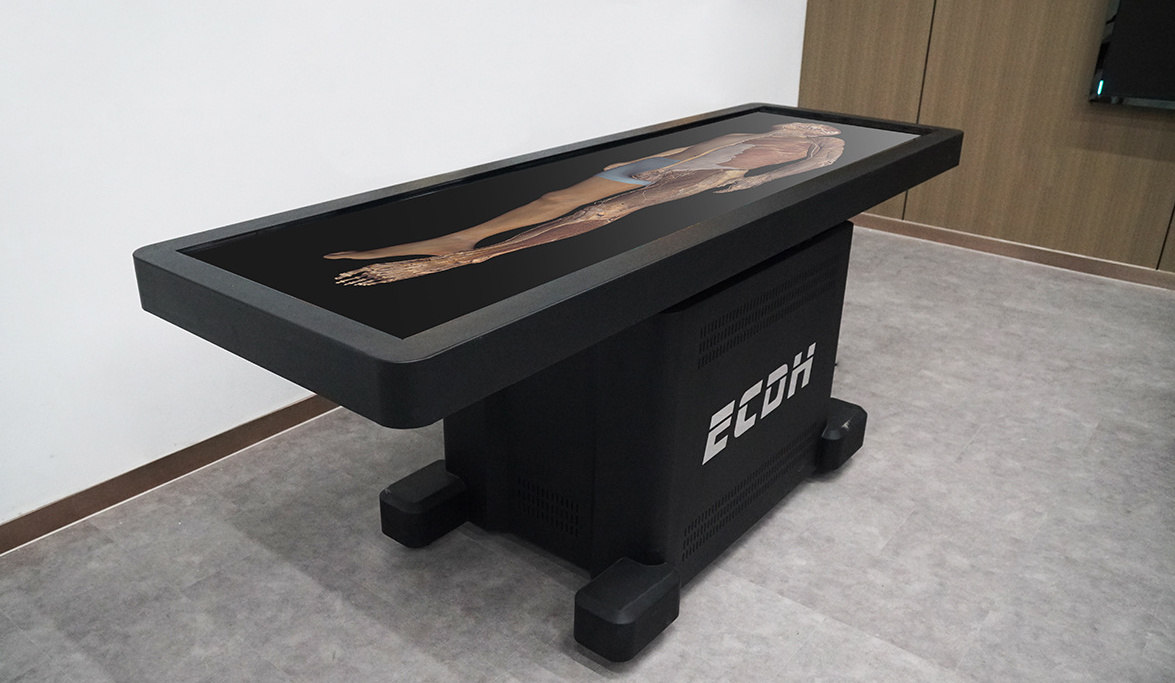
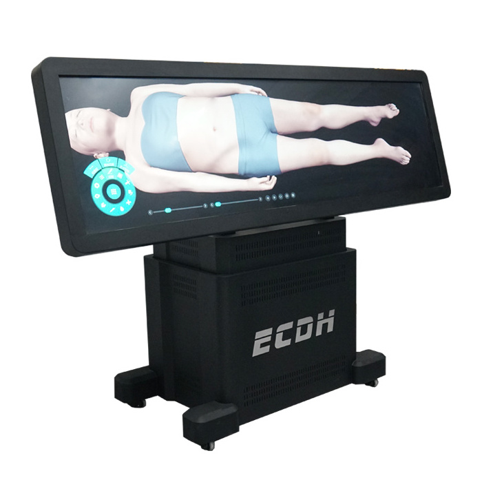
The digihuman virtual dissection table is reconstructed from the real data of continuous tomography of the Chinese human body with no organic resection and no missing, which truly restores the organ structure of the human body. It can be rotated arbitrarily, zoomed in and out of the local structure, and can be observed in multiple directions; cut arbitrarily and view the fault; it can display each structure and arbitrarily added and segmented local organs according to the level, which can well express the adjacent relationship of the organs. It can meet a variety of teaching and training needs.
1. System features
|
Types |
The amount of data |
|
Male |
Image of whole body cross section specimen: The total number of cross-faults is 17,571 Layer thickness 0.1mm Resolution 13700*6340 Split structure 1318 structures |
|
Female |
Image of whole body cross section specimen: The total number of layers of the transverse fault is 16141 Layer thickness 0.1mm Resolution size: 12000*5700 |
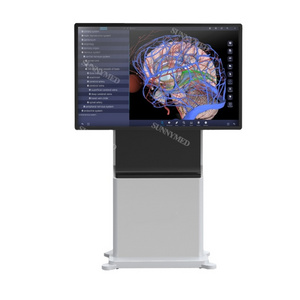
The "High-definition Digital Human Virtual Anatomy System" realizes the real-time rendering of ultra-high-precision data of human body structure through technical processing of ultra-high-precision human tomographic sequence images. The touch operation of the dissection table can realize the section and observation of the human body structure in different directions and levels at any angle, integrate the knowledge of human anatomy, regional anatomy, and sectional anatomy, and help the observation by constructing a virtual anatomical combination model The authors established the concept of three-dimensional spatial structure, effectively improved the quality of education, and reduced the shortage of physical specimens.
The system has five modules: s ystematic anatomy, regional anatomy, sectional anatomy, anatomy micro course and autonomous learning.
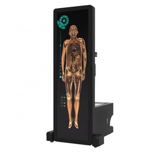
In this system, human structures can display in 3D, which can be enlarged, reduced and rotated at any angle.
In the system, the anatomical structure in each fault of the transverse, sagittal and coronal sections shall be circled and marked to facilitate viewing the position and range of each anatomical structure in the fault, and must be associated with the 3D human body. Click any structure position of the 3D or fault, and other areas will respond synchronously.
The preset position function is set in the system to facilitate the teacher to establish magnetic stickers according to the teaching content, and quickly find the set 3D human body structure during the teaching. Each preset position magnetic sticker contains the histological slice pictures of the corresponding anatomical structure.
The structures can be displayed separately, stripped, recovered, dyed, transparent , searched, painted and so on.
Morphological section
28. The morphological section library contains histological sections and pathological sections, which are classified according to tissues and parts. Sections can be quickly searched, classified and screened.
29. All sections are panoramic slices, which can be enlarged by4X/10X/20X/40X and infinitely enlarged.
30. All sections in the library are equipped with section introduction, including specimen source, staining method, specimen description, knowledge point labeling, etc., and can be quickly reset.
31. The total number of sections more than 2000.
Medical image case database:
32. The images include CT images and MRI images. The images are sequence images, which can be browsed. Each case contains basic information, main complaint, imaging findings, imaging diagnosis, etc.
33. The images contain DICOM information, support window adjustment view and MPR reconstruction, the sectional, sagittal and coronal images, and view the images from any angle.
34. Key position of images can be measured and marked.
35. All cases can be reconstructed, which is convenient to view the 3D structure generated by the image, and the generated 3D structure can be cut and observed arbitrarily.
36. Image can be imported and viewed in DICOM format.
5. Exhibition mode Full touch operation display.
Packaging & Shipping
Selling Units: Single item
Single package size: X50X250 cm
Single gross weight: 500.000 kg
Package Type: Standard for Export
Our Services
1. The Hardware Quality Assurance of the Products stipulated in this Contract shall be 36 months after the arrival of the Goods at the Port of Destination and during the period damage caused by the buyer's considerations shall be the buyer's own responsibility; Software can be upgraded free of charge